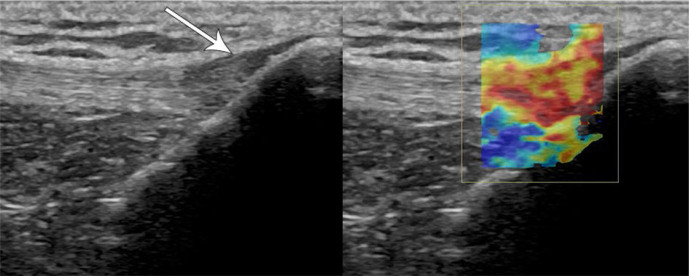
Fig. 16.
65-year-old female with anterior knee pain. Longitudinal grayscale and transient elastography US images of the distal patellar tendon show focal hypoechogenicity and thickening (arrow) with corresponding increased stiffness denoted by overlying color map (increasing stiffness from blue to red), consistent with focal patellar tendinosis

