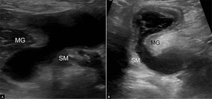
Fig. 17.
A. 19-year-old male with Baker’s cyst. Transverse grayscale US image of the posterior knee shows bilobed anechoic fluid collection located between the semimembranosus (SM) and medial gastrocnemius (MG) tendons, consistent with a Baker’s cyst. Note anisotropy of the SM relative to MG tendon, not to be confused with debris. B. 85-year-old female with complex Baker’s cyst. Transverse grayscale US image shows thick-walled collection with internal septations and debris with otherwise typical location and appearance

