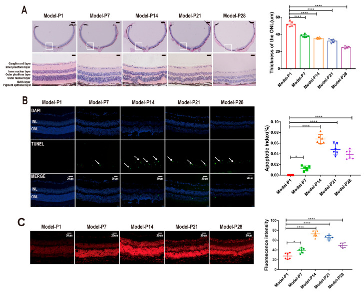
Figure 2.
Morphological damage in the RD model. (A) The retinal morphology was damaged profoundly over time in the RD model. (B) The number of apoptotic cells in the RD model increased significantly. The apoptosis index peaked at P14. The white arrows represent TUNEL-positive cells. (C) The DHE staining showed that ROS accumulated in the retina of the RD model (one-way ANOVA multiple comparisons were analyzed; ns, * p < 0.05, **** p < 0.0001 for differences compared with P1; n = 6).

