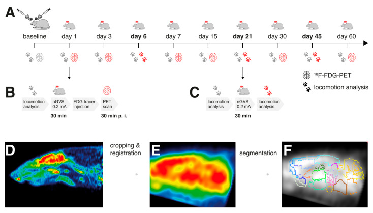
Figure 1.
Experimental protocol and processing of brain scans: (A) 18F-FDG-PET and locomotion analysis were performed before (baseline) and post-bilateral labyrinthectomy (BL) on 9 sequential days encompassing a total period of 60 days. A 30 min long treatment with noisy galvanic vestibular stimulation at 0.2 mA (nGVS; N = 8) vs. sham (N = 9) was applied on each assessment day post-BL. (B) On days 1, 3, 7, 15, 30, and 60 post-BL, immediate stimulation effects on regional brain activation were tested by whole-brain 18F-FDG-PET imaging directly following nGVS treatment. A concomitant locomotion analysis before nGVS treatment was performed on each of these days to assess potential long-term effects of stimulation. (C) Immediate effects of nGVS on locomotion performance were assessed on days 6, 21, and 45 post-BL. (D–F) Post-processing of PET serial scans: Images were acquired, reconstructed, cropped, registered, filtered, normalized, and segmented into 26 brain regions. Subsequently, activity levels for each brain region normalized to the whole brain were obtained.

