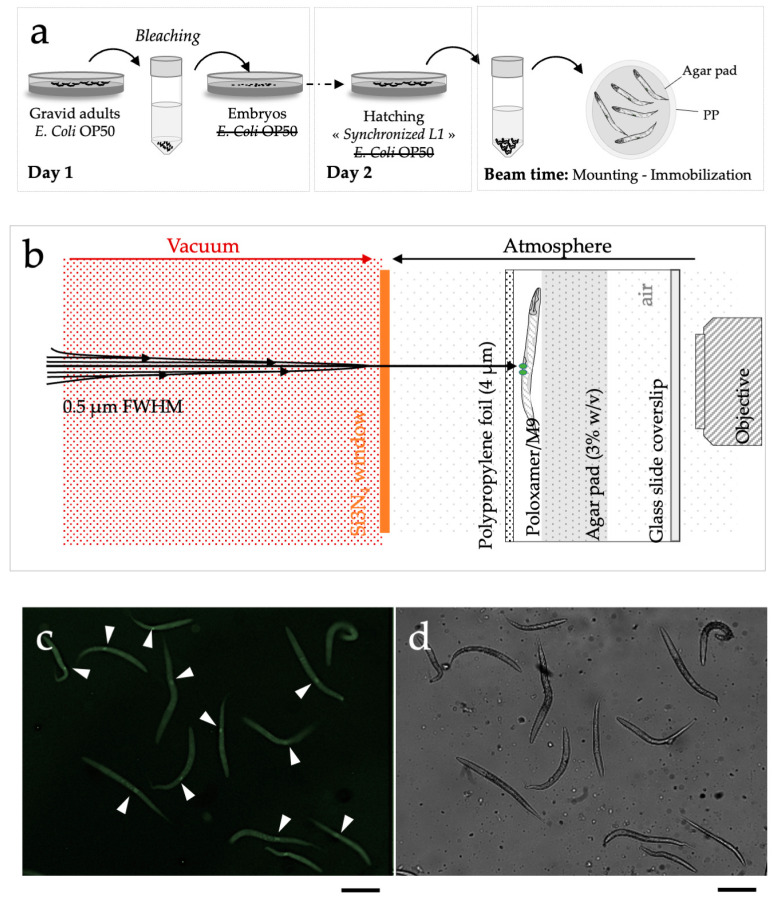
Figure 2.
Schematic representation of the different steps needed for micro-irradiation of C. elegans larvae (early L1 stage). (a) Preparation of large populations of early C. elegans L1 larvae by bleaching in the absence of E. coli OP50. (b) Detailed scheme of the microbeam end- station. 30 min before irradiation, an aliquot ~2 µL was directly deposited on a sterile 4 μm thick polypropylene (PP) foil and immediately covered with an afresh agar pad to maintain immobilized worms in a thin layer of medium. To prevent desiccation and contamination, the dish was closed with a glass side coverslip. Z2-Z3 nuclei were targeted using online fluorescence microscopy. The beam was positioned on the targeted cell. (c) PCN1::GFP detection in Z2-Z3 cells in synchronized early L1 using on-line fluorescence microscopy as obtained in real experimental conditions. White arrows indicate Z2-Z3 GFP-positive cells in synchronized early L1 larvae. (d) Synchronized early L1 larvae visualized using phase contrast imaging. scale bar: 100 µm.

