Figure 3.
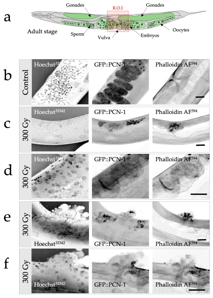
Radiation-induced alterations of gonadal and vulval development in C. elegans following selective and targeted irradiation analyzed by confocal microscopy. (a) Schematic representation of the gonads and vulva in an adult worm (control). The region of interest (ROI) is depicted with the red square. (b) Confocal imaging of the ROI of an adult worm. Nuclei (DNA) are visualized using Hoechst33342 (left column), in utero fertilized embryos are revealed thanks to the GFP reporter (middle column), and the vulvar muscles with their typical cross-sectional profile are detected with the help of phalloidin (actin fibers; right column). (c–f). Confocal imaging of the ROI of structural alterations detected in irradiated worms (300 Gy). Several configurations were observed. (c) Gonadal and vulval agenesis, absence of gonadal and vulval development with a limited actin fibrillar network, no embryos detected, absence of the typical cross-sectional profile of the vulval muscles. (d) Gonadal agenesis and vulvar developmental anomalies. Evidence of tissue disorganization illustrated by the actin fibrillar network and absence of gonads. (e,f) Gonadal agenesis and abnormal vulval eversion. Tissular disorganization, cell nuclei proliferation and disorganization, alteration of the actin fibrillar network, and evidence of abnormal vulvar eversion and agenesis. Scale bar: 10 µm.

