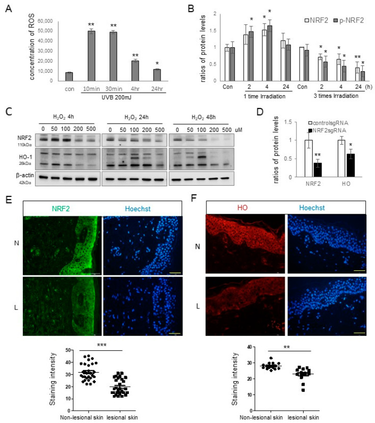
Figure 1.
NRF2 downregulation in the lesional epidermis of patients with melasma. (A) Reactive oxygen species concentrations at various time points in primary cultured keratinocytes following UVB irradiation. (B) Western blot analyses showing NRF2 protein level ratios after single and repeated UVB radiation. (C) Western blot analyses illustrating NRF2 and HO-1 protein level ratios over time in primary cultured normal human keratinocytes treated with different concentrations of H2O2. (D) Western blot analyses presenting HO-1 protein level ratios in cultured human keratinocytes with or without NRF2 knockdown. β-actin served as the internal control for the Western blot analysis. The data are presented as means ± SD from four or eight independent experiments. (E,F) Representative immunofluorescence staining using anti-NRF2 (E) and anti-HO-1 antibodies (F) in the lesional (L) and non-lesional (N) epidermis of patients with melasma. The nuclei were counterstained with Hoechst 33258 (scale bar = 0.05 mm), and the intensities were quantified using ImageJ software 1.54d. * p < 0.05, ** p < 0.01, *** p < 0.001.

