Figure 2.
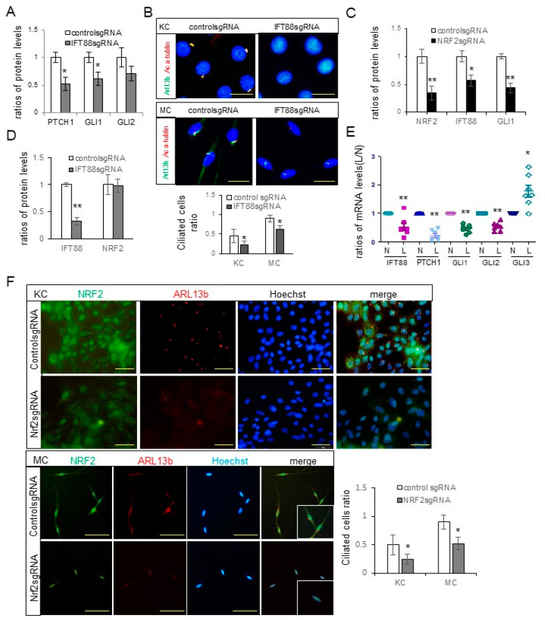
Downregulation of NRF2 led to reduced expressions of IFT88 and Hh signaling molecules involved in ciliogenesis. (A) Western blot analyses depicting the ratios of PTCH, GLI1, and GLI2 levels in cultured keratinocytes subjected to IFT88 knockdown. (B) Confocal microscopy images illustrating primary cilia stained with anti-acetylated α-tubulin (Ac α-tubulin) and/or ARL13b antibodies in cultured human keratinocytes and melanocytes, with or without IFT88 knockdown (bar = 0.05 mm). The ciliated cell ratios were calculated by counting the number of ciliated cells among 30 cells. (C,D) Western blot analyses showing the ratios of NRF2, IFT88, and/or GLI1 levels in cultured keratinocytes with knockdowns of NRF2 (C) or IFT88 (D). (E) Real-time PCR results displaying the ratios of IFT88, PTCH1, and GLI1-3 mRNA levels in lesional compared to non-lesional skin specimens (seven sets) from melasma patients with downregulated NRF2. (F) Representative immunofluorescence staining for primary cilia using anti-NRF2 and anti-ARL13b antibodies in primary cultured human keratinocytes and melanocytes with or without NRF2 knockdown (scale bar = 0.05 mm). β-actin and GAPDH served as internal controls for the Western blot analysis and real-time PCR, respectively. The data are presented as means ± SD from four independent experiments. * p < 0.05, ** p < 0.01.

