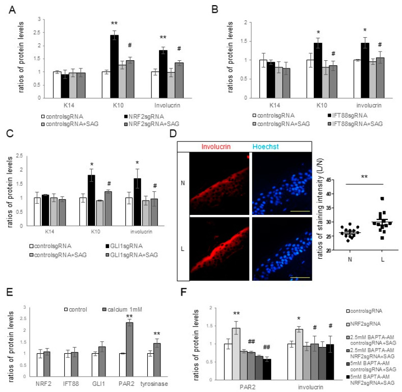
Figure 4.
Effects of NRF2, IFT88, and GLI1 knockdowns on keratinocyte differentiation and subsequent hyperpigmentation. (A–C) Western blot analyses illustrating the relative levels of K14, K10, and involucrin in cultured keratinocytes with or without knockdowns of NRF2 (A), IFT88 (B), or GLI1 (C) in the absence and presence of SAG. (D) Representative immunofluorescence staining using anti-involucrin antibodies (B) in the lesional (L) and non-lesional (N) epidermis of seven patients with melasma. The nuclei were counterstained with Hoechst 33258 (bar = 0.05 mm), and the intensities were measured using ImageJ software 1.54d. (E,F) Western blot analyses for the ratios of tyrosinase levels in keratinocyte–melanocyte cocultures and the ratios of PAR2, K10, involucrin, NRF2, IFT88, and/or GLI1 levels in cultured keratinocytes, including those treated with calcium (E) and keratinocytes with or without NRF2 knockdown in the absence and presence of Bapta-AM (F). β-actin was used as an internal control for the Western blot analysis. The data are presented as the means ± SD from four independent experiments. * p < 0.05, ** p < 0.01 vs. control sgRNA; # p < 0.05, ## p < 0.01 vs. without SAG treatment.

