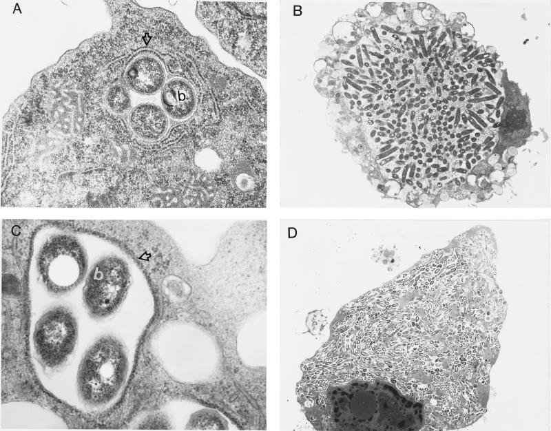
FIG. 2.
Transmission electron micrographs of H. vermiformis (A and B) and WI-26 type I human alveolar epithelial cells (C and D) infected with L. pneumophila AA100 at 4 h (A and C) and 12 h (B and D) postinfection. The open arrows in panels A and C indicate RER-surrounded phagosomes, while the b’s indicate bacteria. Note that the whole cell (B and D) becomes heavily infected with numerous bacteria (a few hundred to a thousand) by 18 h postinfection. Magnifications, ×20,400 (A), ×3,400 (B), ×27,200 (C), ×1,700 (D).

