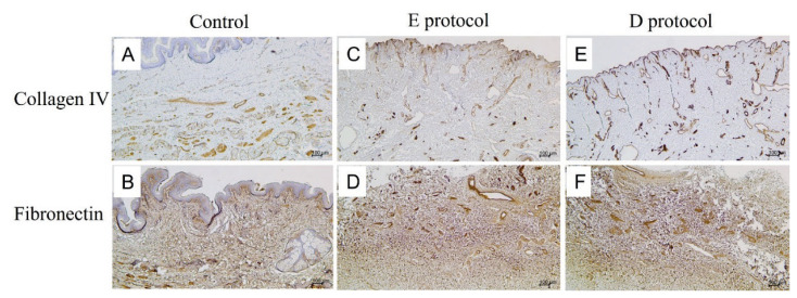
Fig. 4.
Immunostaining for collagen IV and fibronectin in native controls (A, B) and decellularized specimens (C–F). The native control stained for collagen IV (A) displays positivity mainly around blood vessels, and epithelial lining as the integral component of basal laminae. Both the E protocol (C) and D protocol (E) of decellularization preserved collagen IV as evidenced by the absence of any significant changes in collagen IV positivity after cell removal. Fibronectin in the native control (B) is widely distributed in the dermal layer as a crucial component of the ECM. In both the E protocol (D) and D protocol (F) of decellularization, fibronectin positivity is similar in distribution compared to native controls.

