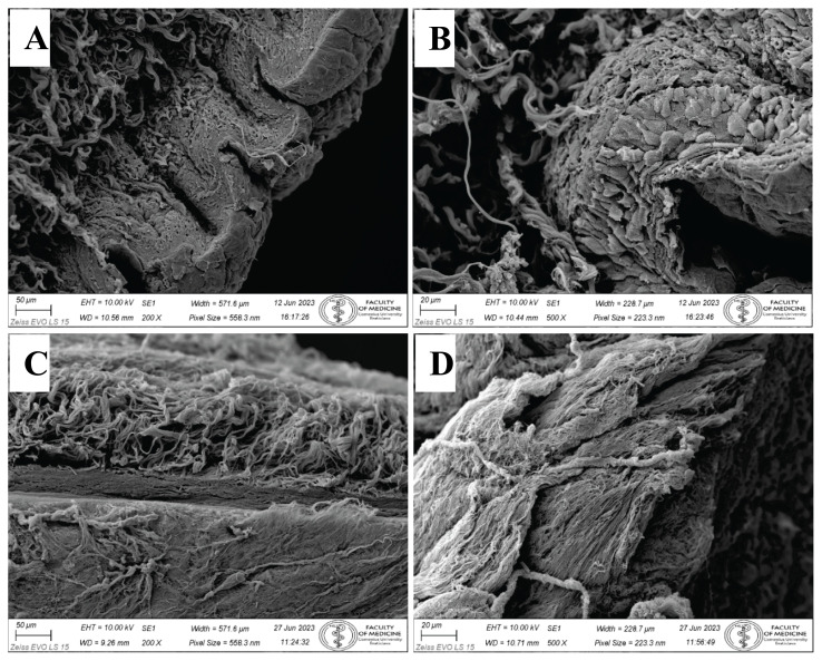
Fig. 5.
Topography of control foreskin before the decellularization process by SEM. A) Stratified squamous keratinized epithelium covers the foreskin surface. Collagen fibers in underlying collagenous connective tissue form bundles in the dermis. Mag. 200x. B) Detail of all layers in the stratified squamous keratinized epithelium. Individual epithelial cells are closely packed and bound by intercellular connections. Mag. 500x. Topography of foreskin after the decellularization process by SEM. C) No epithelium is present on the sample surface. Only collagenous connective tissue of lamina propria remains. Collagen fibers in deep layers represent the foreskin reticular layer of the dermis. Mag. 200x. D) Naked surface of foreskin dermal papillae. Collagen fibers are evident. No epithelium is preserved. Mag. 500x.

