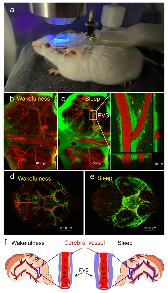
Figure 1.
The changes in activity of the brain waste removal system (BWRS) during sleep and wakefulness: (a) photo of real-time multiphoton monitoring of BWRS in non-anesthetized mouse under EEG control. (b,c) Representative images of real-time multiphoton microscopy of fluorescein isothiocyanate-dextran (FITCD, green) distribution in perivascular spaces (PVSs) surrounding the cerebral vessels filled with Evans Blue dye (EBD, red) after its injection into the right lateral ventricle in awake (b) and sleeping (c) male mouse under EEG control. During wakefulness, PVSs are not filled with FITCD and appear empty. However, during sleep, PVSs are completely filled with FITCD. (d,e) Representative ex vivo confocal images of FITCD distribution in the brain after its injection into the right lateral ventricle in awake (d) and sleeping (e) mice. The intensity of fluorescent signal from FITCD is higher in sleeping vs. waking brain. (f) Schematic illustration of changes in PVS size during wakefulness and sleep.

