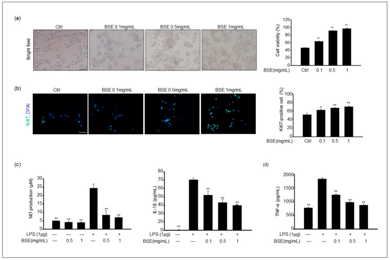
Figure 2.
Effect of 70% EtOH broccoli sprout extract (BSE) on RAW 264.7 cell viability and production of NO, interleukin-1β (IL-1β), and tumor necrosis factor-α (TNF-α) in lipopolysaccharide (LPS)-stimulated RAW 264.7 cells. (a) The cell was treated with 0–1 mg/mL BSE for 24 h. The corresponding graph represents the percentage of cell viability. (b) BSE-treated cells were stained with Ki67 antibody, and the corresponding graph shows the percentage of Ki67-positive cells. Values of * p < 0.05 and ** p < 0.01 were considered statistically significant compared to the controls. Scale bar = 100 μm. (c) Cells were pretreated with BSE for 1 h and stimulated with 1 μg/mL LPS for 24 h. The levels of NO in cultured medium were quantified with Griess reagent. Values of ** p < 0.01 were considered statistically significant compared to cells treated with LPS only. (d) The levels of IL-1β and TNF-α in RAW 264.7 cells were analyzed via an ELISA. Data are shown as the mean ± standard deviation of the mean (SD) of three independent experiments. Values of ** p < 0.01 were considered statistically significant compared to cells treated with LPS only.

