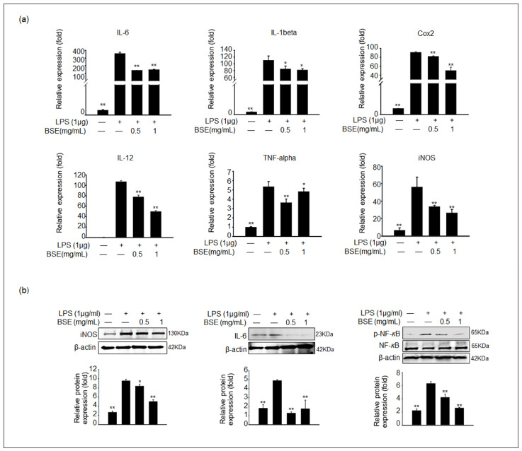
Figure 3.
Effect of BSE on the expression of proinflammatory cytokines in LPS-stimulated RAW 264.7 cells. Cells were pretreated with BSE for 1 h before LPS treatment for 24 h. (a) The mRNA expression of IL-6, IL-1β, COX-2, IL-12, TNF-α, and iNOS was analyzed via qPCR (n = 5). Values represent the mean ± SEM. (b) The protein expression levels of iNOS, IL-6, and phosphorylated nuclear factor-κB (phospho-NF-κB) in BSE- and LPS-treated RAW cells were analyzed via immunoblotting. The density of protein bands was normalized to that of β-actin or the inactive form using Image J software Version 1.53t. Data are shown as the mean ± SD of three independent experiments. All values of * p < 0.05 and ** p < 0.01 were considered statistically significant compared to cells treated with LPS only.

