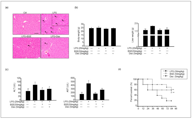
Figure 4.
BSE ameliorated the damage and survival rate of liver tissue after an LPS challenge. (a) Images show hematoxylin and eosin staining of liver tissue from each experimental group: control, LPS only, BSE administration with LPS injection, and both dexamethasone (Dex)- and LPS-injected. Black arrows and circles indicate the infiltration of immune cells. Scale bar = 50 μm. (b) Body and liver weight from each experimental group. (c) Serum alanine aminotransferase (ALT) and aspartate aminotransferase (AST) levels were measured in experimental groups. (d) Mice were orally injected with BSE (daily) for 2 weeks. Treatment of mice with i.p. injection of Dex (5 mg/kg) 24 h before injection with 30 mg/kg LPS. On the final day, LPS was injected into the control, BSE, and Dex groups. Survival rates of mice were determined 84 h postinjection of LPS. Data are shown as the mean ± SEM of three independent experiments. Values of * p < 0.05, ** p < 0.01 were considered statistically significant compared to LPS-only groups.

