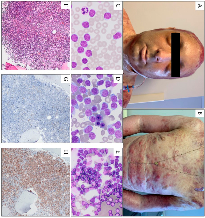
Figure 3.
Pink-red papules, nodules, and tumors scattered on the face torso, and upper limbs in a 32-year-old male with T-PLL. A confluent erythema with a hemorrhagic reaction is also visible in the area of the scalp. Generalized skin edema, scattered petechiae, and conjunctivitis are also visible (A,B). Peripheral blood smear (1000× magnification) (C) and bone marrow smear (1000× magnification) (D) and fine-needle skin aspiration smear (1000× magnification) (E) show numerous atypical prolymphocytes. In bone marrow trephine, a homogenous infiltrate by CD3-positive, TdT-negative prolymphocytes is seen; hematoxylin and eosin staining image under 100× magnification (F) and in immunohistochemistry for TDT (100× magnification, (G)), and CD3 (100× magnification, (H)).

