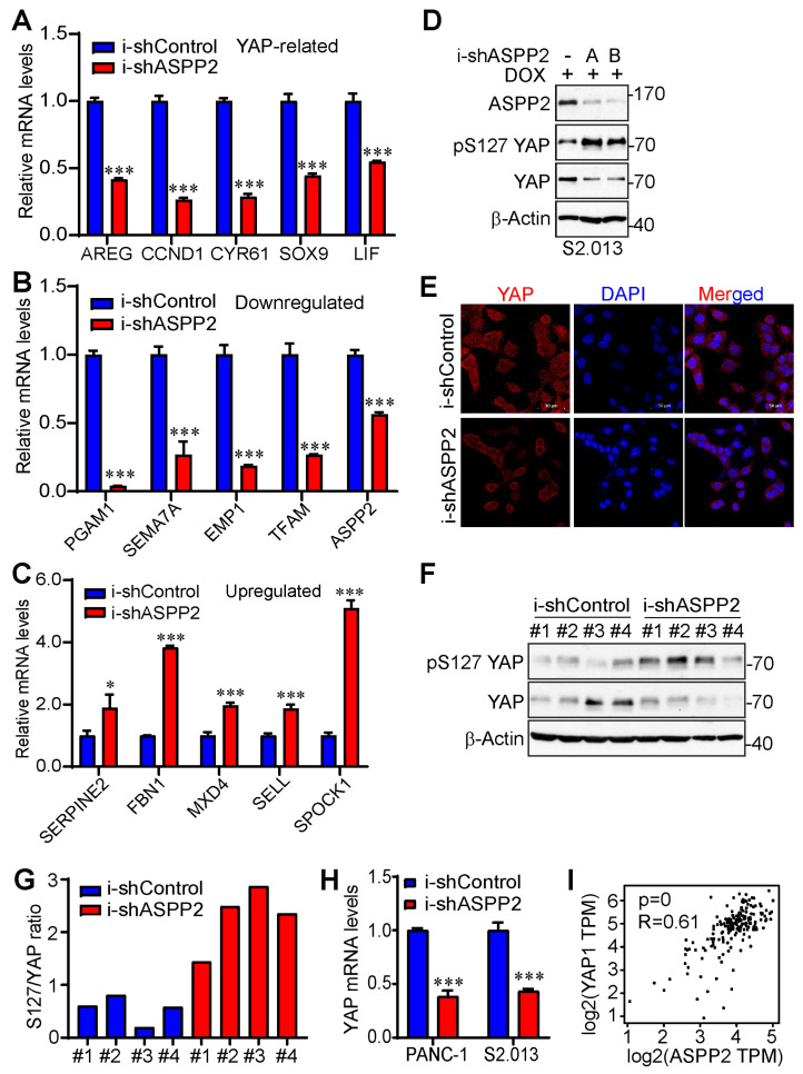
Figure 6.
ASPP2 promotes YAP activity. (A–C) RT-PCR validating YAP-related genes (A), downregulated genes (B), and upregulated genes (C) by ASPP2 depletion from the top differentially expressed gene list. *: p < 0.05; ***: p < 0.001 (Student’s t test). Data are expressed as the mean ± SD of three independent experiments. (D) Western blot analysis showing increased phosphorylation of YAP at S127 and decreased YAP total proteins in ASPP2-depleted S2.013 cells. (E) Representative immunofluorescence staining images illustrating cytosolic retention of YAP in ASPP2 knockdown S2.013 cells. YAP was stained by an Alexa Fluor® 647 anti-YAP antibody indicated in red. The nucleus was stained by DAPI and is marked by the blue color. Scale bar: 50 µm. (F,G) YAP activity was reduced in ASPP2 knockdown tumors. Tumor samples (#1–#4) were probed with the indicated antibodies (F). Quantification was performed using ImageJ (G). (H) RT-PCR showing the relative mRNA expression of YAP in ASPP2 knockdown and control cell lines. ***: p = 0.000048 in PANC-1 and p = 0.00017 in S2.013 (Student’s t test). Data are expressed as the mean ± SD from three independent experiments. (I) Correlation analysis of ASPP2 and YAP mRNA expression based on the TCGA database using GEPIA2. The uncropped bolts are shown in Supplementary Materials.

