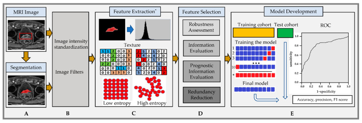
Figure 3.
Radiomic feature analysis workflow: (A) Segmentation of tumor (bounding box in red) on postcontrast T1w images acquired from an 82-year-old male patient with bladder cancer extending to the prostate with a VIRADS score of 5. (B) Image standardization, a preprocessing step. (C) Feature extraction* from the region of interest with the histogram and textural image feature representation with low and high entropy. (D) Feature selection approaches. (E) Model development. Note: * Extracted features should be compatible with the Image Biomarker Standardization Initiative (IBSI) [57].

