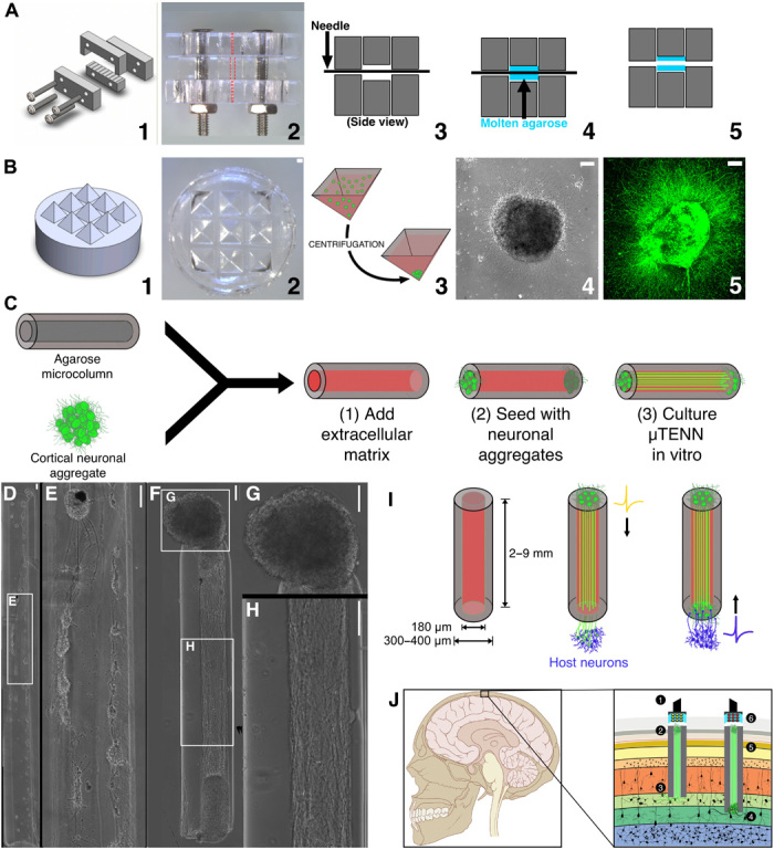
Fig. 1. Aggregate μTENN fabrication and living electrode concept.

μTENNs comprise a hydrogel microcolumn, living neuronal aggregates, and an extracellular matrix lumen. (A) 1: A customizable acrylic mold for generating microcolumns. 2: Top view of the mold dashed lines indicate the outer diameter (OD; middle) and the inner diameter (ID; top and bottom). 3: Needles of the desired inner diameter are inserted into the mold. 4: Microcolumns are cast in agarose (blue). 5: Microcolumns are removed after needle removal and mold disassembly. (B) 1: A three-dimensional (3D) printed mold for square pyramidal wells. 2: Pyramidal wells cast in polydimethylsiloxane (PDMS). 3: Dissociated neurons (green) centrifuged in the wells to form spheroidal aggregates. 4: Phase image of an aggregate 24 hours after plating. 5: Confocal reconstruction of aggregate at 72 hours, labeled with green fluorescent protein (GFP). (C) Microcolumns (gray) are filled with an extracellular collagen-laminin matrix (red). Neuronal aggregates are then placed at the microcolumn terminal(s) and grown in vitro. (D) Early-generation μTENNs fabricated with dissociated neurons yielded limited control over the final network structure (E). Aggregate μTENNs (F) exhibit robust axonal growth and more controllable architecture, with discrete regions of cell bodies (G) and neuritic projections (H). (I) Left: Current μTENN dimensions for implantable living electrodes. Middle: Unidirectional μTENNs synapse host neurons (purple) to relay external inputs to targeted cortical regions. Right: Host neurons synapse bidirectional μTENNs, relaying activity from host cortex for monitoring via the dorsal aggregate. (J) Optogenetically active μTENNs as transplantable input/output channels. Inputs: An LED array (1) optically stimulates a unidirectional, channelrhodopsin-positive μTENN (2) to activate layer IV neurons (3). Outputs: Layer V neurons (4) synapse a bidirectional μTENN (5); relayed neuronal activity is recorded by a photodiode array on the brain surface (6). Scale bars, 100 μm. Photo credit: Dayo O. Adewole, University of Pennsylvania.
