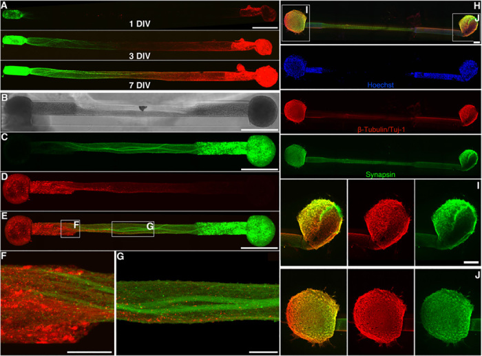
Fig. 3. Aggregate μTENN architecture.

Bidirectional μTENNs were labeled with GFP (green) and mCherry (red) to observe aggregate-specific axonal growth and structure in vitro. (A) Confocal reconstructions of a bidirectional, GFP/mCherry-labeled μTENN at 1, 3, and 7 DIV. (B) Phase image of a bidirectional, GFP/mCherry-labeled μTENN at 5 DIV. (C to E) Confocal reconstruction of the μTENN from (B) at 7 DIV, with insets showing axons from each aggregate making contact with the opposing population (F) and growing along each other (G) in the microcolumn lumen. (H) Confocal reconstruction of a representative bidirectional μTENN at 10 DIV, immunolabeled for cell nuclei (Hoechst, blue), axons (β-tubulin III/Tuj-1, red), and synapses (synapsin, green). Cell bodies are localized to the microcolumn terminals with axonal tracts spanning the distance. Insets in (H) refer to callout boxes (I) and (J) showing aggregate zoom-ins of synapses, axonal networks, and their overlay. Scale bars, 500 μm (A to C and E); 100 μm (F and G); and 200 μm (H and I).
