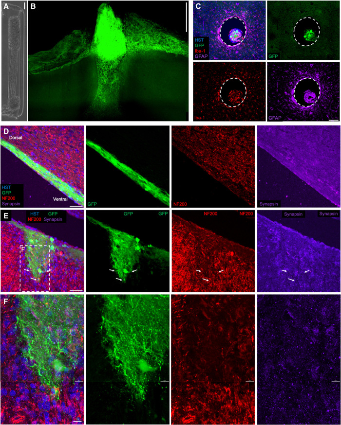
Fig. 6. Living electrode survival and integration in vivo.
(A) Phase image of a bidirectional μTENN before implantation; aggregates have been internalized to the microcolumn. (B) Multiphoton image of the μTENN from (F) at 1 month after implant, showing GCaMP+ μTENN neurons and processes within and immediately surrounding the construct. At 1 month, the dorsal aggregate had descended into the microcolumn, suggesting that externalized aggregates may be required to maintain a cohesive neuronal population at the surface. (C) Axial view of implant showing μTENN neurons/axons in the lumen at 1 month after implant. To visualize the extent of the inflammation response at 1 month following delivery, sections orthogonal to the implant site were stained for microglia/macrophages (Iba-1, red) and astrocytes (GFAP, far red). Minimal host neuroinflammatory response was observed at this time point surrounding the implanted construct. Dashed lines denote the host brain-μTENN interface. (D to F) Longitudinal view of implanted μTENN within the corticothalamic tract at 1 month after implant, with evidence that the construct retained its axonal tracts and overall axosomatic architecture (GFP, green). (E) μTENN neurons and axons (GFP, green) were found projecting ventrally with neurons and neurites interfacing with host tissue at the (deep) ventral end. GFP+ μTENN neurons were visualized in discrete regions with axons extending within the lumen parallel to the cortical-thalamic axis. These findings demonstrate that following stereotaxic microinjection, aggregate μTENNs survive, with neurite extension and integration out to at least 1 month in vivo. Arrows denote μTENN neurites penetrating the host brain and putative synapse formation, with dashed inset representing zoom-in (F). Scale bars, 100 μm (A to C); 50 μm (D and E); and 10 μm (F). HST, Hoechst.

