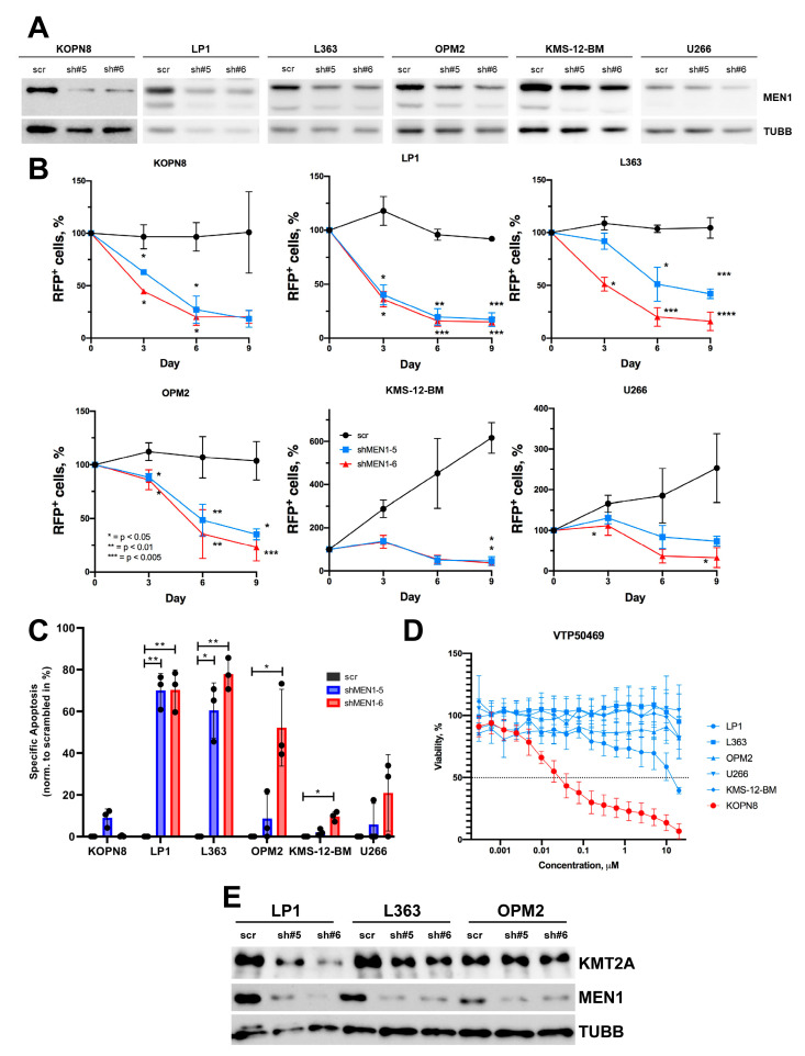
Figure 3.
ShRNA-dependent MEN1 knockdown decreases fitness and induces apoptosis in MM cell lines (A–C) MM cell lines were transduced with shRNAs (sh#5 or sh#6) or control RFP+. (A) On day 4, RFP+ cells were sorted and MEN1 expression was measured by immunoblot. TUBB was used as loading control. (B) MEN1 knockdown decreases fitness of MM cell lines. Starting from day 4 after transduction (day 0) the percentage of the RFP+ cells was measured by flow cytometry. The data are shown as percent of RFP+ cells (mean ± SD, N = 3). (C) MEN1 knockdown induces apoptosis in MM cell lines. RFP+ cells were sorted on day 3 after transduction and incubated under normal conditions for 72 h. Apoptosis was measured by Annexin V/7-AAD staining. The data are shown as specific apoptosis as described in the legend to Figure 2. * = p < 0.05, ** = p < 0.01, *** = p < 0.005, **** = p < 0.001 (D) MM cell lines are not sensitive to VTP50469. The sensitivity was measured by 6 days MTT metabolic test. The MLLr B-ALL cell line KOPN8 was used as a positive control. The data are shown as percentage to control treated with vehicle DMSO (mean ± SD, N = 3). (E) MEN1 KD downregulates KMT2A expression in LP1 and L363 cell lines. The transduced cells were sorted and analyzed by immunoblot as described in (A–C). The representative one of three independent experiments yielding similar results is shown.

