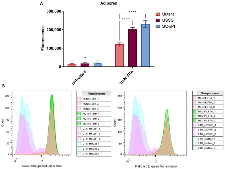
Figure 8.
Mitochondrial hypermethylation induces lipid accumulation under lipid stress, without further lipid peroxidation. (A) Quantification of lipid droplets in both untreated and 24 h treated with 1 mM FFA cell lines (MCviPI mutant (mutant), MsssI (MSSSI), and MCviPI) with Adipored fluorescent staining. Data are shown as mean ± s.d.; n = 3 independent biological replicates (ns = not significant; **** p < 0.0001, two-way ANOVA with Tukey’s correction for multiple comparisons). (B) Lipid peroxidation quantification with the Image-iT Lipid Peroxidation kit using flow cytometry. The lipid peroxidation reagent is a ratiometric probe, and the signal is detected on a flow cytometer with 488 nm laser excitation and fluorescence emission measured at 530/30 nm and 532 nm laser excitation and fluorescence emission measured at 585/42 nm. The data are represented as the ratio of red/green fluorescence intensities. Ratios are lower (indicating greener signal) in cells treated with cumene hydroperoxide (positive control; CTR), but there is no difference between ratios of the MCviPI mutant (Mutant) and MCviPI cell line, both untreated and treated with 1 mM FFA for 24 h (n = 3 independent technical replicates).

