Figure 1.
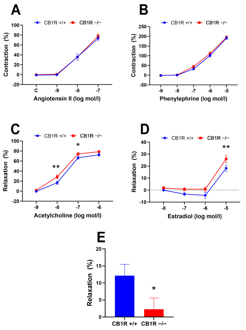
Contraction–relaxation vascular responses of aortic segments of wild-type (CB1R+/+) and CB1R knockout (CB1R−/−) female mice. Panel (A). Dose–response contraction curves to angiotensin II in wild-type (n = 14–25 rings from 9 animals) and in CB1R knockout (18–26 segments from 9 animals). Panel (B). Dose–response contraction curves to phenylephrine in CB1R+/+ (n = 28 segments from 9 animals) and in CB1R−/− (n = 27 segments from 9 animals). Panel (C). Dose–response relaxation curves to acetylcholine in CB1R+/+ (n = 26 segments from 9 animals) and in CB1R−/− (n = 27 segments from 8 animals). Panel (D). Dose–response relaxation curves to estradiol in CB1R+/+ (n = 8–10 segments from 8 animals) and CB1R−/− (n = 7–10 segments from 10 animals). Panel (E). Relaxation response of aortic segments to the CB1R agonist WIN 55,212-2 (10µM) in CB1R+/+ (n = 5) and CB1R−/− (n = 6) female mice. Mean ± SEM values. p < 0.05 values were considered significant. *: p < 0.05 and **: p < 0.01 between wild-type (CB1R+/+) and CB1R knockout (CB1R−/−) groups in two-way ANOVA with a Bonferroni post hoc test and unpaired t-test (panel E) (Contraction data were normalized to KCl contraction, relaxation data were calculated as percent values of precontraction level).

