Figure 2.
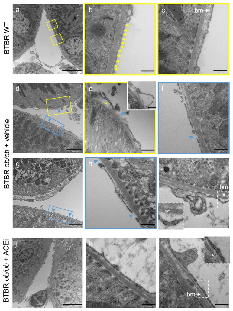
The peritubular capillary ultrastructure is altered in BTBR ob/ob mice and ameliorated by ACEi treatment. Representative TEM microscopy images of peritubular capillary ultrastructure in BTBR WT mice and in diabetic BTBR ob/ob mice received vehicle or treated with the ACEi lisinopril. (a–c) Overview and details of a peritubular capillary in BTBR WT: normally fenestrated endothelium with diaphragms (arrows, b) and basement membrane (c). (d–i) Peritubular capillaries from vehicle treated BTBR ob/ob mice showing focal separation and delamination of endothelial cell from the basement membrane (asterisks, (d,e); insert in (e)), with lucent areas and amorphous material, denuded basement membrane (arrowheads, (d–h)) or foci of basement membrane thickening (bm, (i)). (j–l) Peritubular capillary from ACEi-treated mouse with normal-appearing structure (j). Images at high magnification from other capillaries showing endothelial cells covering their basement membrane ((k,l), inset). Bar = 5000 nm (a,j); 2000 nm (d,g) and 500 nm (b,c,e,f,h,i,k,l).

