Figure 3.
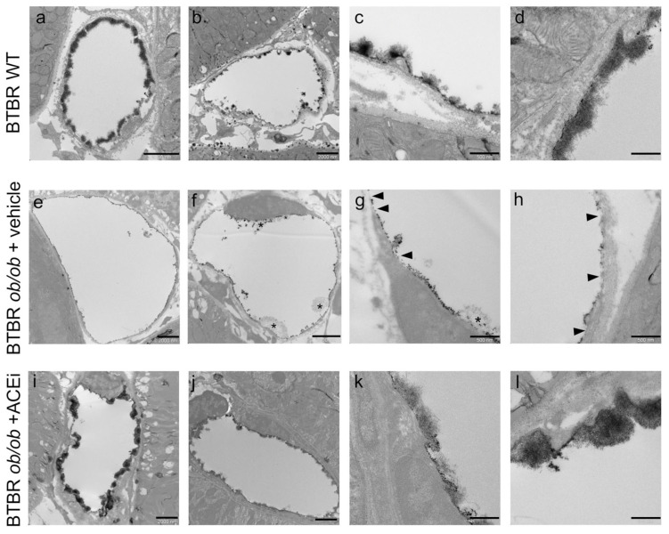
The endothelial glycocalyx of peritubular capillaries is altered in diabetic mice and preserved by ACEi treatment. Representative TEM images of lanthanum-stained endothelial glycocalyx of peritubular capillaries in BTBR WT mice and in diabetic BTBR ob/ob mice received vehicle or treated with the ACEi lisinopril. (a–d) The endothelial glycocalyx of peritubular capillaries in BTBR WT showing a dense layer covering the luminal surface of the capillary with a moss-like pattern well visible at high magnification (c,d). (e–h) The endothelial glycocalyx in diabetic mice appears granular and discontinuous with areas of loss or reduced coverage ((g,h) arrowheads) and the occasional presence of loose network ((f,g) asterisks). (i–l) The endothelial glycocalyx is maintained by lisinopril treatment showing a pattern similar to that observed in WT mice. Bar = 2000 nm (a,b,e,f,i,j) and 500 nm (c,d,g,h,k,l).

