Figure 5.
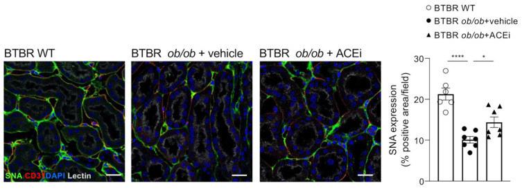
Staining for sialic acid is reduced in peritubular capillaries of diabetic mice and improved by ACEi treatment. Representative images and quantification of sialic acid (SNA, green) immunofluorescence staining in the renal interstitium of BTBR WT mice and diabetic BTBR ob/ob mice received vehicle or treated with the ACEi lisinopril. Data are mean ± SEM. * p < 0.05, **** p < 0.0001. Cell nuclei (DAPI, in blue), renal structures (lectin, pseudocolor). Bar = 20 µm.

