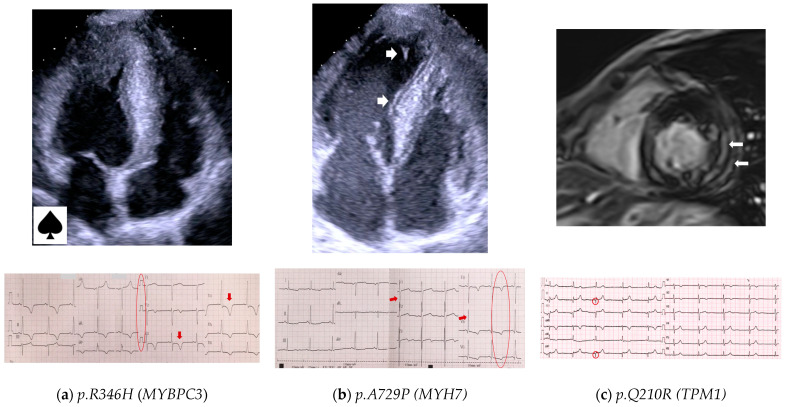
Figure 2.
Cardiac imaging and ECG of HCM patients carrying the rare recurrent variants presented in ≥2 families in our study. (a) A 34-year-old male (42P) carrying the p.R346H variant in MYBPC3; Echo shows apical HCM with LV cavity in the shape of an ace of spades; ECG shows giant negative T waves (arrows) in lateral leads (Yamaguchi syndrome). (b) A 45-year-old female (34P), carrying the p.A729P variant in MYH7; Echo shows asymmetric septal LV hypertrophy and LV hypertrabeculation (arrows); ECG shows LV hypertrophy (Sokolow–Lyon index 45 mm pointed by arrows), and negative T waves (circle) in lateral leads. (c) A 38-year-old female (39Sa) carrying the p.Q210R variant in TPM1; CMR shows asymmetric LV hypertrophy with LGE (arrows); ECG shows Q-waves in leads II, aVF (circles).

