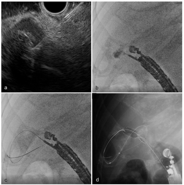
Figure 3.
Techniques of endoscopic ultrasound-guided gallbladder drainage. (a) The gallbladder was detected using endoscopic ultrasound and punctured using a 19 G fine aspiration needle, while avoiding conspicuous blood vessels. (b) After adequate aspiration of infected bile, the contrast agent was administered through the needle. (c) After deep insertion of a guidewire into the gallbladder, the puncture tract was dilated using a dilator along the inserted guidewire. (d) A fully covered dumbbell-type metal stent was placed from the gallbladder to the gastrointestinal tract.

