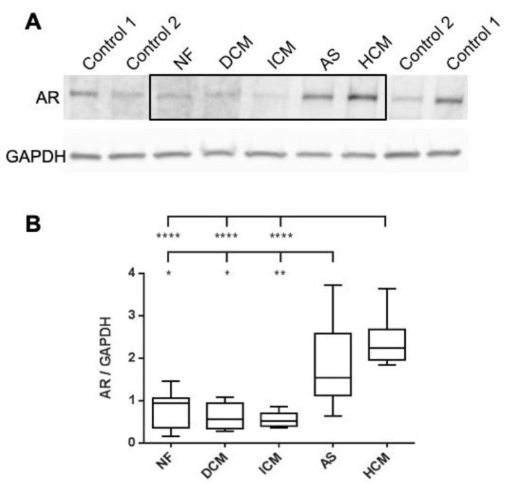
Figure 1.
Western Blot Analysis of AR Protein Levels in Various Cardiac Conditions using the AR-Antibody EPR1535. (A) Representative Western blot demonstrating full-length AR detection at 110 kDa and GAPDH detection at 37 kDa. (B) Box and whisker plot illustrating the summarized statistics of normalized AR signals for each cardiac condition. Sample details are as follows: NF (n = 6), DCM (n = 7), ICM (n = 7), AS (n = 6), HCM (n = 7). Disease group comparisons were conducted using the unpaired t-test. Groups with significant variations in AR expression are marked with respective p-values (* = p < 0.05, ** = p < 0.01, **** = p < 0.0001). Abbreviations: AR = Androgen receptor; AS = Valvular aortic stenosis; GAPDH = Glyceraldehyde-3-phosphate dehydrogenase (housekeeping gene); NF = Nonfailing; DCM = Dilated cardiomyopathy; ICM = Ischemic cardiomyopathy; HCM = Hypertrophic cardiomyopathy.

