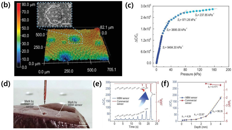
Figure 6.
(a) The fabrication diagrams of pollen-inspired ionic gel based high-sensitive pressure sensor. (b) The 3D topography of the micro-cone surface structured PDMS template. (c) Capacitance change upon a pressure. (d) Penetration marks when identifying needle-tissue contact using commercial and MBM sensors. (e) Real-time monitoring of needle penetration comparison with commercial sensors. (f) The sensitivity upon a penetration depth of the MBM and commercial sensor [32].


