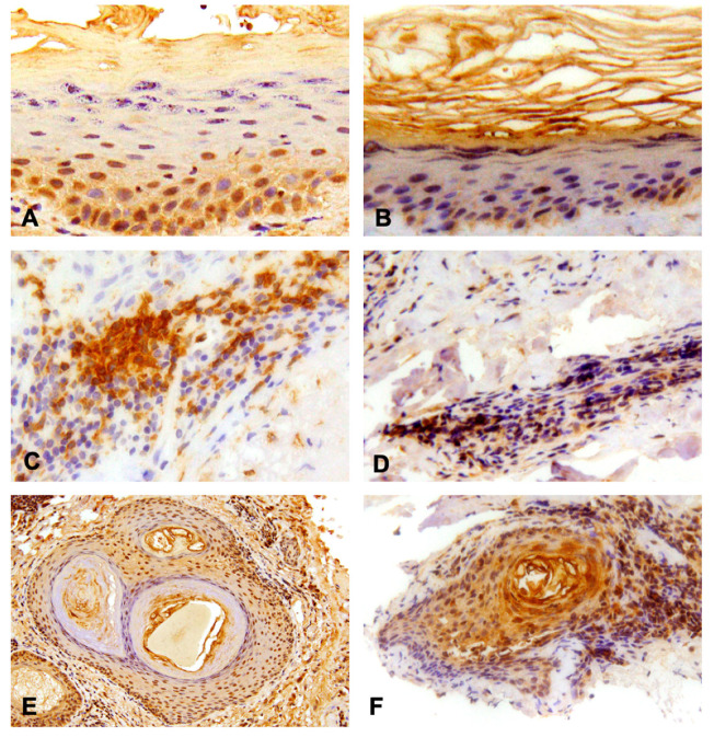
Figure 2.
Results of the immunohistochemistry analysis. (A) Positive expression of TLR-8 in granular layer of the epidermis, 40× magnification; (B) Positive expression of TLR-9 in the spinal layer of the epidermis and partially in the granular one, 20× magnification; (C) Dermal infiltration of lymphocyte TLR-8 (intensively positive), 40× magnification; (D) Dermal infiltration of lymphocyte TLR-9 (mildly positive), 20× magnification; (E) TLR-8 and (F) -9 expression in the context of hair follicles, 10× and 20× magnification respectively.

