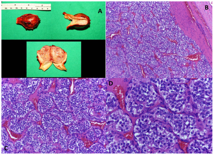
Figure 3.
(A) Macroscopic aspect of carotid paraganglioma. (B) Paraganglioma, peripheral fibrous capsule, and nests of tumor cells separated by conjunctival-vascular septa; H.E. ob. 100×. (C) Paraganglioma, relatively regular nests of tumor cells separated by conjunctival-vascular septa; H.E. ob. 200×. (D) Paraganglioma, round tumor cells with central, regular nuclei and peripheral cytoplasm that is clear or pale eosinophilic and finely granular; H.E. ob. 400×.

