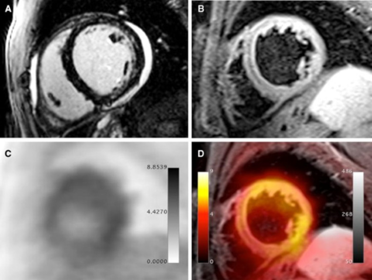
Fig. 5.
PET/MRI examination in a 32-year-old male patient presenting with dyspnea, mild ventricular dysfunction (51% LFEV), and a history of recent systemic viral disease. A shows patchy intramyocardial late gadolinium enhancement in the lateral and inferior wall as well as pericardial effusion. B shows significantly increased T2 signal in the lateral wall representing myocardial edema. C (PET) and D (fusion between T2-weighted MR image and PET) show diffusely increased FDG uptake in the lateral, anterolateral, and inferolateral wall. Histopathological assessment after endomyocardial biopsy showed acute myocarditis with lymphocytic infiltration and moderate myocyte apoptosis. The patient demonstrated elevated levels of C-reactive protein (4.1 mg/dl) as well as elevated myocardiocytolysis serum markers (Troponin-I: 0.42 ng/ml). PCR and immunohistochemical analysis did not detect specific infectious agents such as viruses, bacteria, or fungi.
Reprinted with permission of Springer from [50]

