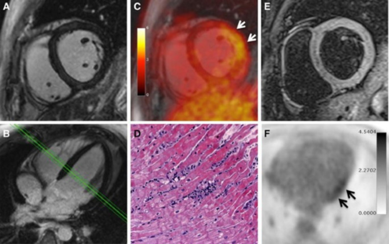
Fig. 6.
PET/MRI examination in a 30-year-old male patient presenting with chest pain, dyspnea, palpitations, severely limited exercise capacity, mild ventricular dysfunction (54% LVEF), and mild ECG abnormalities (T-wave flattening in II, III, aVF, and V6). The patient demonstrated neither elevated levels of C-reactive protein nor elevated myocardiocytolysis serum markers. LGE images (A, B) did not reveal any signs of myocardial necrosis. PET images (C, F) demonstrated focal FDG uptake in the lateral wall. T2-weighted imaging (E) showed mild myocardial edema (T2 ratio: 2.0). Diagnosis of borderline myocarditis was confirmed by histopathological assessment after endomyocardial biopsy demonstrating sparse inflammatory infiltrates but no myocardial necrosis (D). PCR and immunohistochemical analysis did not detect specific infectious agents such as viruses, bacteria, or fungi.
Reprinted with permission of Springer from [50]

