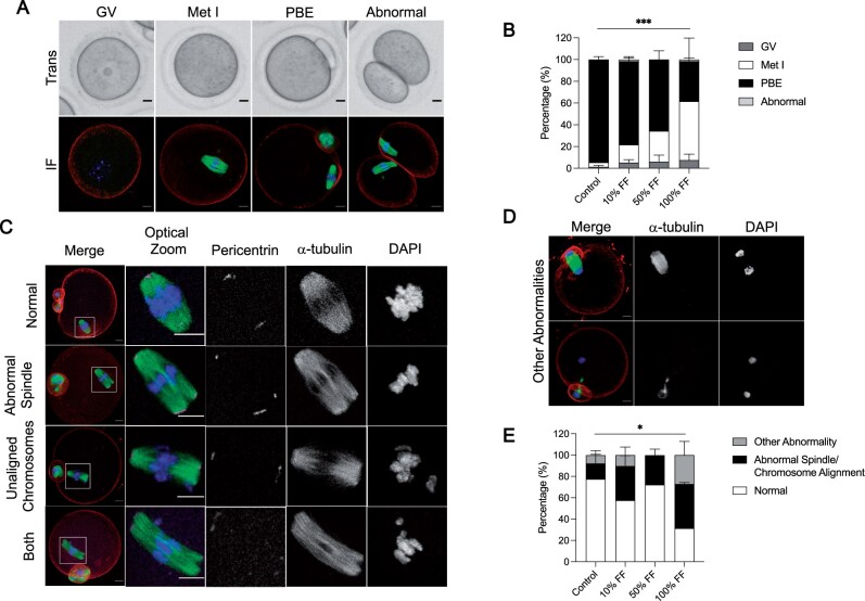
Figure 2.
Follicular fluid (FF) has dose-dependent effects on oocyte meiotic maturation. (A) Representative transmitted light images (top) and immunofluorescence z-projections (bottom) of each meiotic stage. Oocytes with an intact germinal vesicle (GV), metaphase I (Met I) oocytes, eggs that extruded polar bodies (PBE), and oocytes that underwent symmetric cleavage (abnormal) were identified by morphological criteria. Actin (rhodamine phalloidin, red), α-tubulin (green), and DNA (DAPI, blue) were detected by immunocytochemistry. Scale bars = 10 μm for all panels. (B) Graphs of meiotic progression for oocytes matured in different concentrations of FF. Data are shown as mean ± SEM for all panels. χ2 (3, N = 235)=30.84, ***P < 0.0001 for PBE versus no PBE. N = 56–62 oocytes per group across total of three replicates. (C and D) Representative z-projections of abnormal phenotypes of metaphase II eggs matured in different concentrations of FF. Actin (rhodamine phalloidin, red), pericentrin (magenta), α-tubulin (green), and DNA (DAPI, blue) were detected by immunocytochemistry. (E) Quantification of abnormal phenotypes in eggs following maturation in different concentrations of FF. N = 13–43 eggs per group across total of three replicates. χ2 (3, N = 114)=9.051, *P = 0.0286 for normal versus abnormal.

