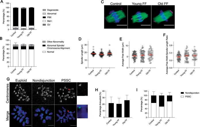
Figure 4.
Follicular fluid (FF) does not impact oocyte meiotic maturation in an age-dependent manner. (A) Graph of meiotic progression for oocytes matured without FF (control), in young FF, or in old FF. Data are shown as the mean±SEM for all panels. N = 234–241 oocytes per group across total of four replicates. (B) Quantification of abnormal phenotypes of metaphase II eggs following maturation without FF (control), in young FF, or in old FF. N = 60–80 eggs per group across total of four replicates. (C) Representative z-projections of metaphase II spindles from eggs following maturation without FF (control), in young FF, or in old FF. Pericentrin (magenta), α-tubulin (green), and DNA (DAPI, blue) were detected by immunocytochemistry. Scale bars = 10 μm. (D–F) Quantification of metaphase II spindle length (D), average pole width (E), and average pole width: spindle length ratio (F) for eggs following maturation without FF (control), in young FF, or in old FF. N = 45–58 eggs per group across total of four replicates. (G) Representative z-projections of metaphase II chromosome spreads from eggs following maturation without FF (control), in young FF, or in old FF. Kinetochores (anti-centromere antibody, green) and DNA (DAPI, blue) were detected by immunocytochemistry. Images labeled with number of kinetochore pairs (top, white). Unpaired kinetochores/chromatids indicated by arrows (red). Scale bars = 5 μm. (H–I) Quantification of aneuploidy rates for eggs following maturation without FF (control), in young FF, or in old FF (H) further divided into nondisjunction or premature separation of sister chromatid (PSSC) errors (I). N = 54–63 eggs per group across total of three replicates.

