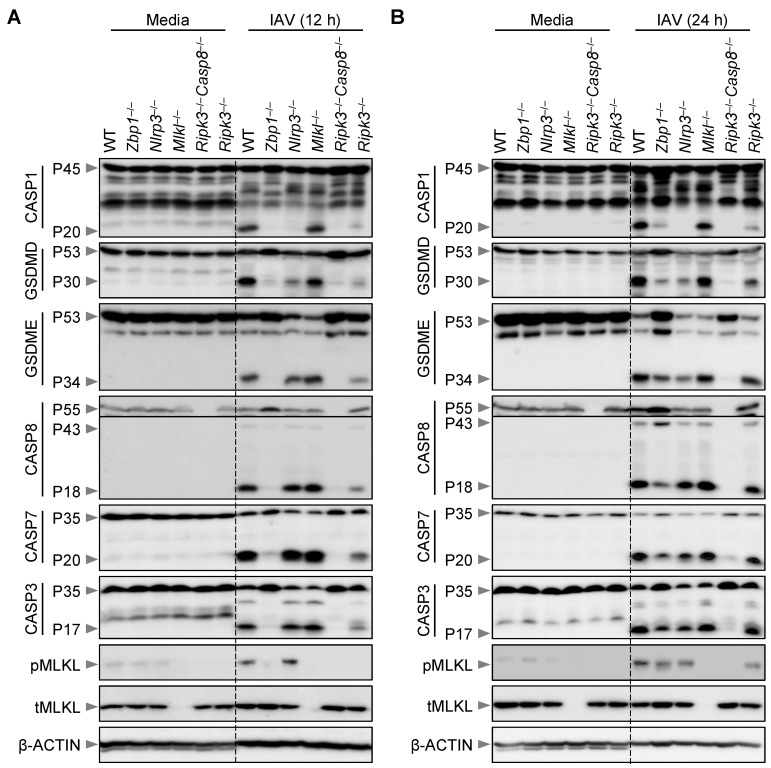
Figure 2.
Loss of MLKL does not protect macrophages from IAV-induced activation of PANoptosis molecules. (A,B) Western blot analysis of PANoptosis in IAV-infected wild-type (WT) and different gene-deficient bone marrow-derived macrophages (BMDMs). Western blot analysis of pro- (P45) and activated (P20) caspase-1 (CASP1), pro- (P53) and activated (P30) gasdermin D (GSDMD), pro- (P53) and activated (P34) gasdermin E (GSDME), pro- (P55) and cleaved (P43 and P18) caspase-8 (CASP8), pro- (P35) and cleaved (P20) caspase-7 (CASP7), pro- (P35) and cleaved (P17) caspase-3 (CASP3), phosphorylated MLKL (pMLKL) and total MLKL (tMLKL) in WT, Zbp1−/−, Nlrp3−/−, Mlkl−/−, Ripk3−/−Casp8−/−, and Ripk3−/− BMDMs at 12 h (A) and 24 h (B) post-infection with IAV. The data are representative of at least three independent experiments (A,B). The uppercase ’P’ in the Western blots represents the molecular weight of the proteins. The lowercase letters ‘p’ and ‘t’ represent the phospho- and total-forms of the proteins, respectively.

