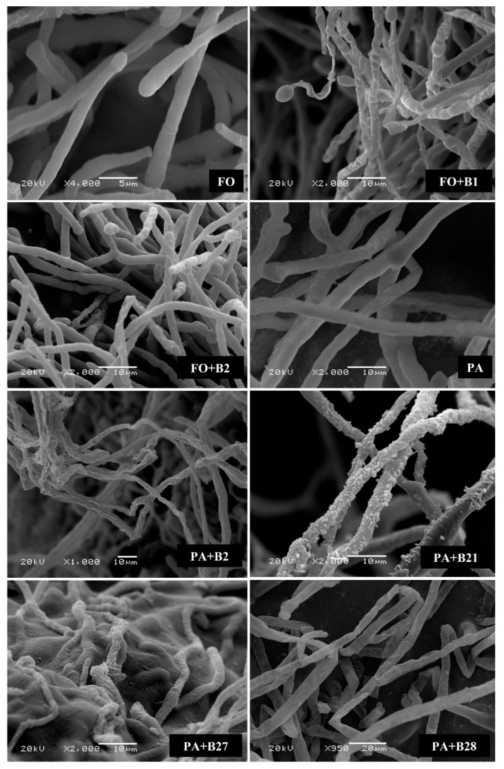
Figure 3.
Antagonistic activity of selected bacterial strains on Fusarium solani (FO) and Pythium aphanidermatum (PA) mycelial morphology depicted using a scanning electron microscope (SEM). Normal patterns of hyphae presented in the control (FS and PA). Distorted mycelial structure, wrinkled or shrunken patterns presented in FO + B1, FO + B2, PA + B2, PA + B21, PA + B27, and PA + B28.

