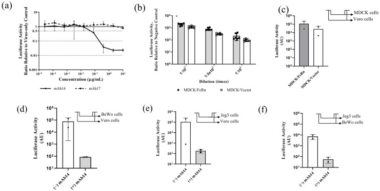
Figure 4.
In vitro applications of nLuc-ZIKV reporter variant. (a) Neutralization of nLuc-ZIKV by monoclonal antibodies in Vero cells. Anti-ZIKV (mAb14, line) and anti-flavivirus (mAb17, broken line) monoclonal antibodies were serially diluted, mixed with nLuc-ZIKV then used to inoculate Vero cells. The infection was quantified using bioluminescence readout and the ratio with the no-antibody controls was plotted as a function of antibody concentration. Each data point represents averages from eight inoculations. Error bars representing standard deviations are shown. (b) Assessing the susceptibility to nLuc-ZIKV infection in cells that overexpress FcRn versus those that do not. MDCK/FcRn cells were more susceptible to nLuc-ZIKV infection than control cells that express an empty vector, recapitulating the findings seen with PRVABC59 strain [12]. Three different dilutions and up to eight replicates per dilution were used for each cell line. Differences are statistically significant by Student t-test (p = 0.036). (c) Evaluating transcytosis of infectious nLuc-ZIKV particles through a semipermeable membrane supporting confluent monolayers of MDCK/FcRn or control cells. Infectious viruses were quantified after transferring the contents of the basal chamber onto Vero cells (shown in the inset panel) and quantifying the infection using bioluminescence readout. Up to six replicates per experiment and cell line were used; the experiment was performed twice. The error bars are outside the lower limit of the y-axis. (d–f) Evaluating transcytosis through placenta trophoblast cells of infectious nLuc-ZIKV particles alone or as immune complexes (IC) with neutralizing antibody. Either BeWo (d) or Jeg-3 (e,f) cells were grown onto semi-permeable trans-well membranes and nLuc-ZIKV alone or as preformed immune complexes with 10 µg/mL anti-ZIKV antibody mAb14 was added inside the trans-well. The contents of the basolateral chamber were transferred onto Vero or BeWo cells as indicated in the inset schematic (top right corner of each panel). Addition of 10 µg/mL mAb14 reduces the infectivity in Vero or BeWo cells. Up to six replicates per experiment/cell line were used; each experiment was performed twice. In panel (e)/(−) IgG column, the error bars are outside the lower limit for y-axis. * p < 0.05.

