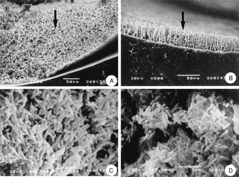
FIG. 3.
Scanning electron micrographs of P. aeruginosa NSM35 growing on catheters. Freeze-dried, freeze-fractured cross sections of colonized all-silicone (A) and hydrogel-coated latex (B) catheters are shown. The arrows indicate the bacterial biofilms colonizing the lumenal surfaces of the catheters. Critical-point-dried preparations showing the surfaces of these biofilms on all-silicone (C) and hydrogel-coated (D) catheters are also shown.

