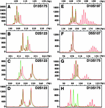
Figure 1.
Type A and Type B microsatellite instability observed in human colorectal cancer. Using genomic DNA samples prepared from cancer and the corresponding normal mucosa, microsatellite sequences, indicated at the right top of each panel, were amplified by PCR with primers differentially labelled with fluorescence, then mixed and run on a same lane in an automated DNA sequencer. The amount of each DNA fragment was quantitatively detected and its size was estimated with accuracy of 1 bp, by standardization with size markers run in each lane. Results representative for each mode of microsatellite instability are shown: red lines, cancer; green lines, normal mucosa; Type A, (A) (IC678), (B) (IC810), (C) (IC721) and (D) (IC793); Type B, (E) (IC790), (F) (IC733), (G) (IC690) and (H) (NoTa). Patient codes in the parentheses correspond to those used in Table 1.

