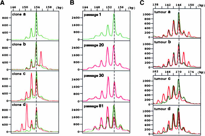
Figure 2.
Microsatellite changes in Msh2−/− mouse embryonic fibroblast (MEF) cells and in tumours that arose in Msh2-knockout mice. (A) More than 20 subclones were isolated from RH95021 (Msh2−/−) MEF cells and microsatellite changes were compared among these subclones. The majority exhibited the same configuration (clone a) and a few deviated from this predominant pattern (clone b–d). In each clone, the pattern of clone A has been superimposed with green lines, to facilitate comparison. Results obtained in D6Mit59 microsatellite are shown. (B) RH95021 cells were continuously cultured and sampled at different passages. Results obtained in D6Mit59 microsatellite at passage 1, 20, 30 and 81 are shown. In passage 81, the initial profile at passage 1 has been superimposed with a green line. (C) Tumours that arose in vivo in Msh2-knockout mice were analysed. Representative results obtained in D7Mit91 (tumours a and b) and D6Mit59 (tumours c and d) microsatellites are shown: red lines, tumour; green lines, the corresponding normal tissue.

