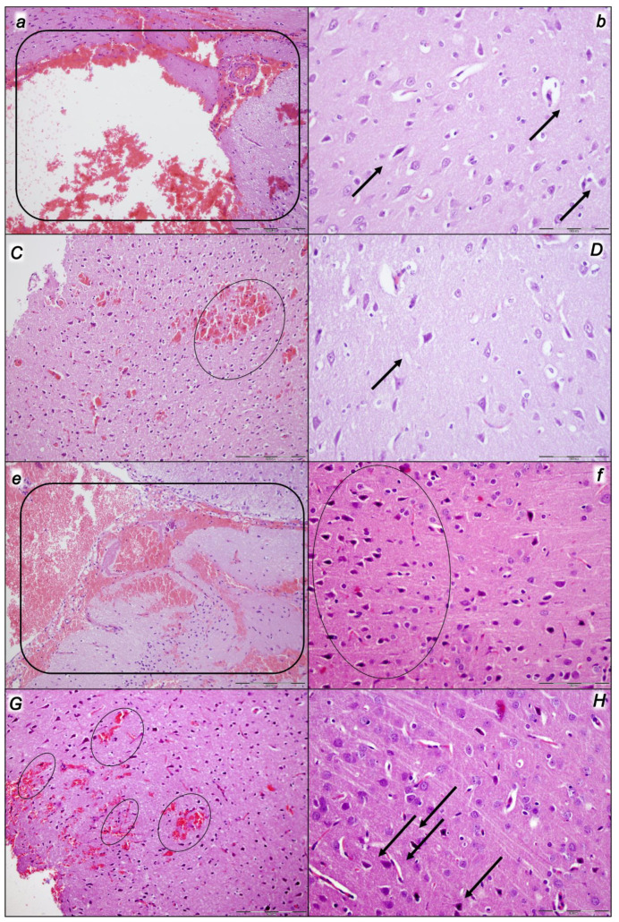
Figure 9.
Neuropathological changes presented in the cerebrum (a,b,C,D,e,f,G,H) in control rats (small italic letters) (a,b,e,f) and BPC 157-treated rats (capital italic letters) (C,D,G,H). Considerable lesions were noted after decompression and reperfusion in rats who were subjected to the intra-abdominal hypertension of 25 mmHg for 60 min (a,b,C,D) (i) or 40 mmHg for 30 min (ii) (e,f,G,H) and sacrificed after the corresponding reperfusion period (60 min (i) (a,b,C,D) or 30 min (ii)) (e,f,G,H) depending on whether they had received (sc) saline (controls) or BPC 157 at 3 min reperfusion times. Commonly, controls presented a pronounced edema and congestion in the brain tissue. In the BPC 157-treated rats (capital italic letters), only mild edema in the brain tissue was found. A focal and deeper neocortical hemorrhage was found in control animals affecting the neocortex, corpus callosum, amygdala, and striatum in the brain tissue (marked areas—(i): (a); (ii): (e)). In the BPC 157 group, only smaller areas of neocortical hemorrhage occurred (marked areas—(i): (C); (ii): (G)). Moderate to severe neurodegenerative changes were presented in the cerebral cortex, along with the karyopyknosis of cortical neurons, in controls (marked areas, black arrows) ((i): (b); (ii): (f)) while BPC 157 rats exhibited, consistently, only rare karyopyknotic cells and mild neurodegenerative changes ((i): (D); (ii): (H)). HE staining; magnification, 200×; scale bar, 200 μm.

