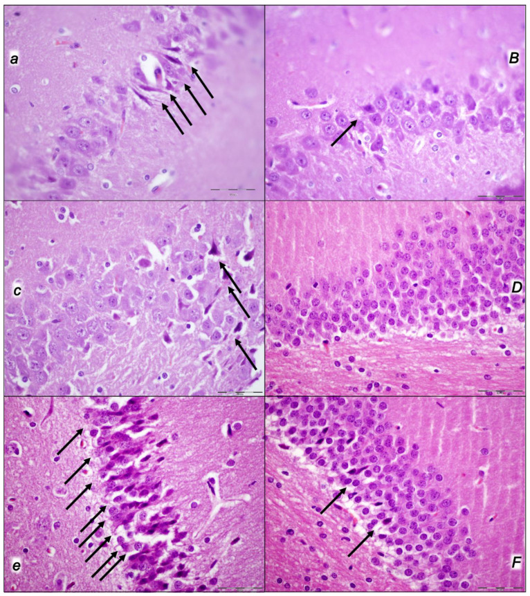
Figure 11.
Neuropathological changes presented in the hippocampus (a,B,c,D,e,F) in control rats (small italic letters) (a,c,e) and BPC 157-treated rats (capital italic letters) (B,D,F). Considerable lesions were noted after decompression and reperfusion in rats who were subjected to the intra-abdominal hypertension of 25 mmHg for 60 min (a,B) (i), 30 mmHg for 30 min (ii) (c,D), or 40 mmHg for 30 min (iii) (e,F) and sacrificed after the corresponding reperfusion period (60 min (i) (a,B), 30 min ((ii) (c,D) and (iii) (e,F))) depending on whether they had received (sc) saline (controls) or BPC 157 at 3 min reperfusion times. Commonly, in the brain tissue, pronounced edema and congestion occurred in controls. In the hippocampus, there were moderate neurodegenerative changes in the control animals. There were karyopyknosis and the degeneration of pyramidal cells of the hippocampus (black arrows). BPC 157 rats exhibited only mild edema in the brain tissue and no or only rare karyopyknotic cells in the hippocampus (black arrows). HE staining; magnification, 600×, scale bar, 50 μm.

