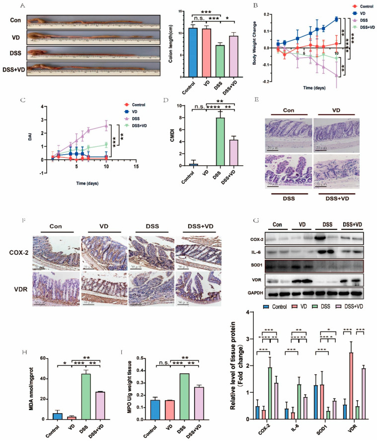
Figure 1.
VD/VDR attenuated DSS-induced UC in mouse model. (A) The length of colon was shortened, thickened, and congested in DSS group, in comparison with DSS + VD group; (B) body weight declined in DSS group compared with DSS + VD group; (C,D) DAI and CMDI scores were notably higher in DSS group than in DSS + VD group; and (E) HE staining of colon was performed after mice were sacrificed. Severe and extensive inflammatory cell infiltration was shown in DSS and DSS + VD groups (scale bar = 50 μm). (F) The expression and distribution of COX-2 and VDR with immunohistochemical stain were shown (scale bar = 50 μm). DSS group showed high expression of COX-2 in comparison with DSS + VD group. (G) The changes in protein levels in different groups were analyzed by Western blot. The contents of COX-2 and IL-6 in DSS group were obviously augmented, whereas the expression of SOD1 was decreased compared with DSS + VD group. (H,I) The levels of MDA and MPO in DSS group were higher than in DSS + VD group. Values shown are means ± SD. * p < 0.05, ** p < 0.01, *** p < 0.001, **** p < 0.0001, and n.s., not significant. (n = 3 in each group).

