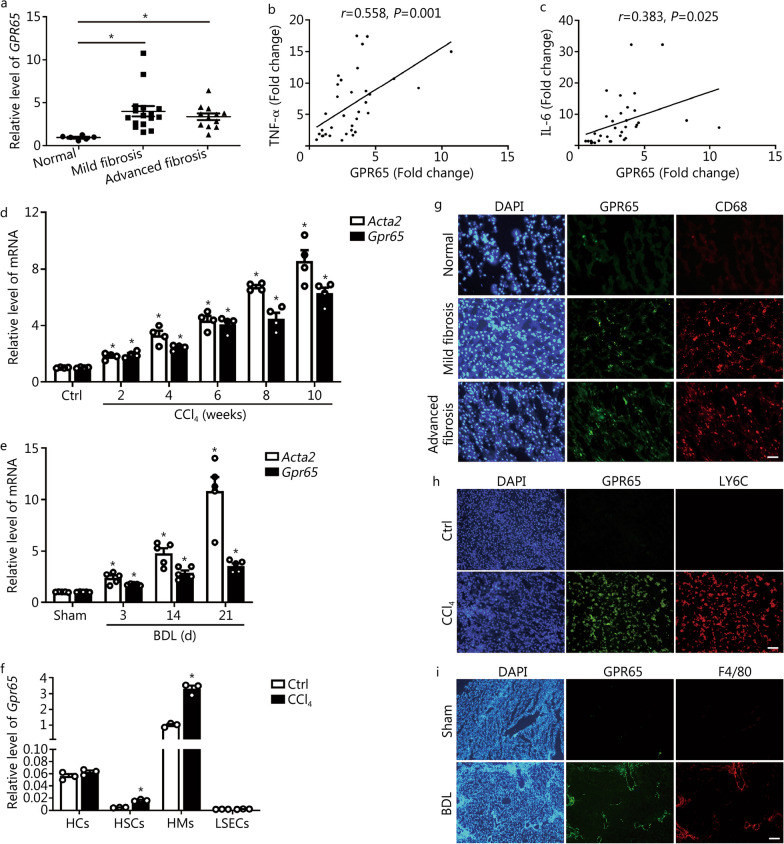
Fig. 1.
GPR65 is overexpressed in mouse and human hepatic fibrosis. a qRT-PCR was used to assess the mRNA level of Gpr65 in liver tissues of human normal controls (n = 6), mild fibrosis (n = 16) and advanced fibrosis (n = 12). The correlations of GPR65, TNF-α (b) and IL-6 (c) were assessed using Pearson correlation analysis, n = 34. d qRT-PCR was used to assess the expression of Acta2 (α-SMA) and Gpr65 in livers from mice with CCl4 treatment for different time points (n = 4). e qRT-PCR was used to assess the expression of Acta2 and Gpr65 in livers from mice treated with BDL for different time points (n = 5). f qRT-PCR was used to assess the expression of Gpr65 in HCs, HSCs, HMs and LSECs that were isolated from livers of mice without or with CCl4 treatment for 8 weeks (n = 3). g Representative IHC-frozen for co-staining GPR65 with macrophage marker CD68 in the liver of human normal controls, mild fibrosis and advanced fibrosis. Scale bar = 50 μm. h Representative IHC-frozen for co-staining GPR65 with macrophage marker LY6C in the livers of mice without or with CCl4 treatment for 8 weeks. Scale bar = 50 μm. i Representative IHC-frozen for co-staining GPR65 with macrophage marker F4/80 in the livers of mice treated without or with BDL for 21 d. Scale bar = 125 μm. *P < 0.05 vs. Normal or Ctrl or Sham. BDL bile duct ligation, CCl4 carbon tetrachloride, Ctrl control, HC hepatocyte, HM hepatic macrophage, HSC hepatic stellate cell, IHC immunohistochemistry, LSEC liver sinusoidal endothelial cell, qRT-PCR quantitative real-time reverse transcription-polymerase chain reaction, TNF-α tumor necrosis factor-α, IL-6 interleukin-6, LY6C lymphocyte antigen 6 complex

