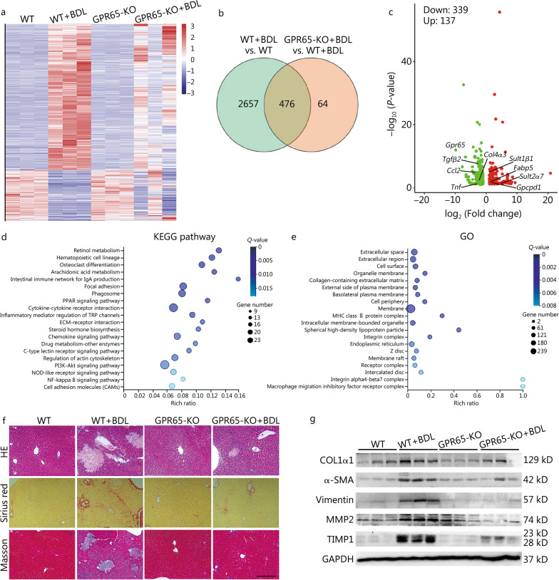
Fig. 2.
Gpr65 deficiency alleviates hepatic fibrosis induced by BDL. a WT and GPR65-KO mice were divided into 4 groups: WT, WT + BDL, GPR65-KO and GPR65-KO + BDL. The significantly differentially expressed mRNAs were displayed by hierarchical cluster analysis: bright red, up-regulation; bright blue, down-regulation. n = 3/group. The Venn diagram (b), volcano map analysis (c), KEGG (d) and GO (e) analysis of differentially expressed mRNAs in WT mice treated with BDL, compared to WT mice, and GPR65-KO mice treated with BDL, compared to WT mice treated with BDL. f Hepatic fibrosis was evaluated by HE staining, Sirius red staining and Masson’s trichrome staining. Scale bar = 400 μm. g Western blotting was used to determine the protein level of COL1α1, α-SMA, vimentin, MMP2 and TIMP1. BDL bile duct ligation, GO Gene Ontology, KEGG Kyoto Encyclopedia of Genes and Genomes, KO knockout, PPAR peroxisome proliferator activated receptor, TRP transient receptor potential, ECM extracellular matrix, NOD nucleotide oligomerization domain, MHC major histocompatibility complex, COL1α1 collagen type I alpha 1, α-SMA α-smooth muscle actin, MMP2 matrix metalloproteinase 2, TIMP1 tissue inhibitor of metalloproteinases 1

