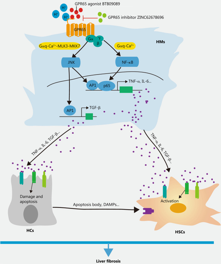
Fig. 8.
Schematic diagram illustrating the function and mechanism of GPR65 in the progression of liver inflammation and fibrosis. Upon liver injury, increased or activated HMs-enriched GPR65 induces up-regulation of TNF-α and IL-6 via the Gαq-Ca2+-JNK/NF-κB pathways, while promoting the expression of TGF-β through the Gαq-Ca2+-MLK3-MKK7-JNK pathway in HMs, thus resulting in the damage of HCs and the activation of HSCs, and subsequently aggravate BDL- and CCl4-induced liver inflammation, injury and fibrosis. In addition, pharmacological inhibition of GPR65 can block this process. BDL bile duct ligation, CCl4 carbon tetrachloride, HC hepatocyte, HM hepatic macrophage, HSC hepatic stellate cell, MLK3 mixed lineage kinase 3, MKK7 mitogen-activated protein kinase kinase 7, NF-κB nuclear factor κB, JNK c-Jun N-terminal kinase, AP1 activator protein 1, TNF-α tumor necrosis factor-α, IL-6 interleukin-6, TGF-β transforming growth factor-β, DAMPs damage-associated molecular patterns

